


Bovine embryonic stem cells yield vascularised, innervated muscle tissue in 15 days
Researchers at EMBL Barcelona have co-induced skeletal muscle, neural and endothelial cells from a single bovine embryonic stem cell line, forming 0.6 mm tissue aggregates within 15 days in serum-free conditions. Published in Nature Communications, the work could simplify cultivated steak manufacturing, but the authors stress that cost, tissue size and blood vessel maturity remain unresolved.
A single cell source for several tissue types
Researchers at EMBL Barcelona have shown that a laboratory-grown line of bovine embryonic stem cells (ESCs) can be guided to form skeletal muscle, neurons and endothelial cells in the same culture. These then organise into three-dimensional, tissue-like aggregates. The study, from Miki Ebisuya, former group leader at EMBL Barcelona and now Humboldt Professor at PoL-TU Dresden, and her team, was published in Nature Communications on 2 September 2026.
For manufacturers, the central issue is the cell source. The paper states that cultivated beef currently relies predominantly on myosatellite cells and mesenchymal stem cells (MSCs) isolated from adult bovine biopsies and bone marrow, and that their proliferation capacity diminishes over long-term culture. Immortalisation can extend that capacity, but the authors note that the genetic modifications involved “may present a challenge in obtaining food authorization”. Adult cells are also restricted in what they can become: myosatellite cells are committed to muscle fate, while the differentiation trajectory of MSCs depends on the tissue from which they were isolated.
Unmodified ESCs, by contrast, proliferate indefinitely and can differentiate into cells of all three germ layers. The authors describe them as more favourable for food production than induced pluripotent stem cells, which need genetic reprogramming. They also note that large-scale culture of bovine ESCs in stirred bioreactors has already been reported. All protocols in the study are serum-free and finish within 15 days. “Our serum-free, rapid co-induction protocols represent a milestone toward self-organizing beef steaks with integrated vasculature and innervation,” the authors wrote in the abstract. Cost and the reliance on Matrigel remain obstacles, discussed below.
The assembly problem in structured beef
Producing structured steak rather than minced meat is the harder task. The paper explains that incorporating several cell types into muscle tissue is key to recreating steaks accurately. For in vitro tissues to keep growing, oxygen and nutrient supply through blood vessels or alternative porous structures is also crucial.
Current manufacturing strategies prepare distinct cell types separately and then assemble them. Techniques include bioprinting purified cells with a bioink, co-culturing cells on edible porous scaffolds and stacking myocyte-laden hydrogel modules. Electrical or mechanical stimuli are often applied to aid muscle maturation and improve texture. The authors say these approaches are promising but add extra manufacturing steps.
“One of the biggest challenges in cultivated meat is reproducing the complexity of real tissue,” said Marina Sanaki-Matsumiya, former postdoctoral fellow at the Ebisuya Group and currently Assistant Professor at the University of Tsukuba in Japan. “Instead of growing each cell type separately and assembling them afterwards, we showed that embryonic stem cells can develop together and self-organise, mimicking real tissue development.”
Stepwise induction through presomitic mesoderm
The team followed the route skeletal muscle takes in the embryo. Bovine ESCs were treated for two days with CHIR (a WNT activator), bFGF, SB431542 (a TGF-beta inhibitor) and DMH1 (a BMP inhibitor) to induce presomitic mesoderm (PSM). Most cells were TBX6-positive on day 2. A HES7 promoter-luciferase reporter showed segmentation clock oscillations with a period of about 4 hours.
The PSM cells were then treated with HGF, IGF and bFGF for four days, followed by HGF and IGF for nine days. Muscle fibres positive for MYOG and sarcomeric α-actinin (SAA) first appeared around day 10 and peaked around day 14. Their average sarcomere length was 2.1 µm, similar to values reported for bovine skeletal muscle in vivo. The authors suggest the roughly 15-day timescale, slightly shorter than in a human protocol, may reflect faster bovine embryonic development.
The fibres showed spontaneous calcium transients, which were suppressed by 10 µM curare, an acetylcholine receptor blocker (p = 5.10E-9; 71 regions of interest from five independent experiments). TUJ1-positive neurons were intermingled with the fibres. Staining for α-bungarotoxin revealed puncta with a rounded, immature morphology, frequently at sites where fibres and neurons were close. The authors interpret these as early neuromuscular junction (NMJ)-like structures. They suggest neuronal stimulation could offer an alternative to the external electrical and mechanical stimuli used in muscle bioengineering.
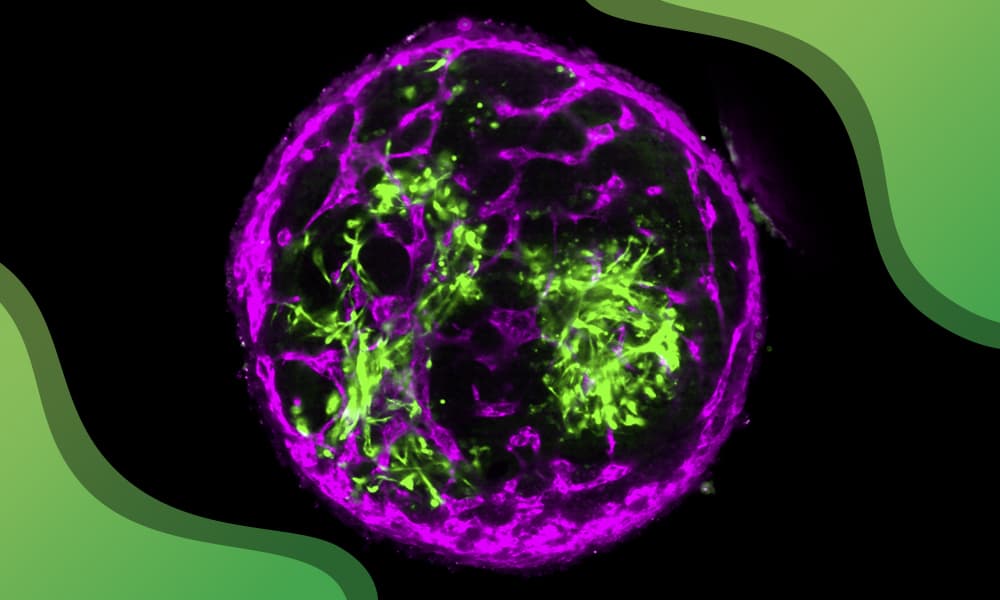
3D bovine tissue containing muscle fibres and blood vessel-like networks.
© Marina Sanaki-Matsumiya/EMBL
Vessel networks and three-dimensional aggregates
To add vasculature, the researchers supplied VEGF and forskolin (a cAMP activator) from day 2 alongside the muscle factors, keeping DMH1 to prevent lateral plate mesoderm differentiation. Elongated VE-cadherin-positive cells appeared around day 8 and peaked around day 14, forming interconnected vessel-like networks. Skeletal muscle domains encased by endothelial cells measured 200-300 µm in diameter on day 15. That is larger than the intercapillary distance of approximately 100 µm in tissues, but the authors describe it as a comparable range.
For the 3D work, 20,000 PSM cells were aggregated on day 2, with 2% Matrigel added to maintain structural integrity. After day 6 the aggregates were cultured on a rotary shaker to aid diffusion of oxygen and nutrients. They were spherical and about 600 µm in diameter, a size that barely changed over the time course, probably reflecting declining proliferation during muscle maturation. Under the muscle and neuron protocol, SAA-positive fibres appeared around day 10, with neural domains forming next to muscle domains. All 57 aggregates exhibited an SAA/DAPI staining ratio of at least 10%. The proportions of muscle and neural domains fell as aggregate size increased.
With the endothelial protocol, VE-cadherin-positive cells emerged around day 10 and formed networks adjacent to the muscle fibres. ZO-1 staining suggested tight junctions. The networks followed a regular pattern and penetrated deeply into the tissue. Without DMH1, the endothelial cells did not form uniform networks and the aggregates were no longer spherical. In these aggregates the muscle and endothelial proportions slightly increased or held constant with size.
Cell composition, practical constraints
Single-cell RNA sequencing of four protocols, each from a single experiment (7,676 to 10,294 cells per sample), identified muscle and neural clusters. An endothelial cluster appeared only under the endothelial protocols. The muscle cells split into skeletal and smooth subclusters, with the 2D protocols yielding more smooth muscle. The authors therefore conclude that the skeletal muscle-rich 3D cultures are more suitable for cultured beef. Adipocyte progenitor, pericyte and osteoblast markers were detected, but mature fat cells were not identified. The authors note that variations such as 2D versus 3D culture can cause unexpected differences in cell type composition.
They list several practical constraints. The protocols were developed with a single bovine ESC line, so independent sources of bovine pluripotent stem cells need testing. “Matrigel, which is derived from mouse carcinoma cells, should be replaced with animal-free alternatives,” they wrote. On cost, they state that “the current costs are commercially unfeasible”: a 0.1 mm³ muscle aggregate derived from 20,000 bovine ESCs costs between 0.39 and 0.99 EUR, with growth factors, chemical inhibitors and Matrigel the main expenses. Mass production of these materials or plant-based alternatives would be needed.
The biological findings carry caveats too. The authors did not observe a clear positive impact of endothelial networks and neurons on muscle cultures, even after 30 days. Whether the networks can support intraluminal perfusion “remains to be seen”.
Next steps towards steak-sized tissue
“The tissue we generated is still very small,” said Ebisuya. “To produce something resembling a steak, we will need much larger tissues with more mature blood vessel networks that can support continued growth.”
The authors identify connecting the endothelial networks to in vitro perfusable vasculature as the next crucial step, since intraluminal flow stimulates lumen formation. They add that perfusion with blood substitutes may improve the flavour of cultured steaks. Co-inducing stromal cells such as pericytes and fibroblasts may aid vessel formation. Maturing the NMJ-like structures may need protocol optimisation and supporting cell types such as Schwann cells. Further refinements may also enable co-induction of fat cells.
The authors see the approach as complementary to engineering methods: co-induced cells can be mixed with a bioink for bioprinting, and muscle aggregates with endothelial networks can serve as building blocks for complex tissue assembly. They report having filed an international patent application related to the work. Beyond food, the authors note that the multi-cell system could help researchers investigate muscle development and tissue engineering. “Despite the remaining challenges, we propose the co-induction of multiple cell types and self-organization of tissues as feasible directions in the production of cultured beef steaks,” the authors concluded.
Reference
Sanaki-Matsumiya, M., Sanaki, Y., Villava, C., et al. (2026). Self-organization of vascularized muscle from bovine embryonic stem cells. Nature Communications, 17, 8975. https://doi.org/10.1038/s41467-026-76569-2